Modern thinking for injuries focuses on one aspect – “I need an image, now!” and “I have to know what is happening, it will help you treat me better.” But is this true? Since the dawn of medicine, when there were no images, they treated based on symptoms. All medicine treats symptoms and analyzes based on symptoms. Medicine is practice. All healthcare is practice to refine and improve diagnosis and treatment. Diagnosis places a patient in a category on how to treat. In some cases this is simple and other cases, very difficult. We as humans are diverse and varied. What causes pain in one person, does not cause pain in another. What is considered a major injury can effect patients differently. The diagnosis category no longer holds weight.
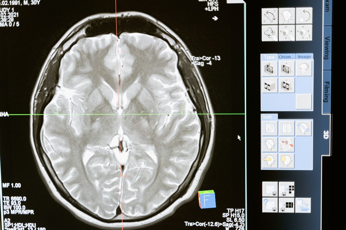
Generally after a simple injury, the most basic imaging will be an x-ray (XR). XR’s help to establish bone, bony abnormalities (bone spurs), pneumonia, some cancers, and objects (2). When there is significant trauma, Emergency Rooms (ER) are very well versed in the imaging needed to determine life or death. Other imaging, like a computerized tomography (CT), show more detail than XRs and provides cross sectional images of bones (even hairline fractures), blood vessels, and soft tissues (1). Contrast can be added to increase the likelihood of finding abnormalities. Magnetic Resonance Imaging (MRI) are considered the Gold Standard for care. MRIs are able to see pretty much everything within the body through radio waves and hydrogen atoms within the body to provide a 3D image (3). Because MRIs are the best imaging, they are often expensive, especially with insurance with costs up to $3000 per image. Some places offer MRI imaging for under $600, cash only, in Springfield, MO.
MRIs are often the first imaging patients want - skip to the chase and get to the right diagnosis. However, because of costs and time, it may not be the appropriate imaging for an injury. This is where doctors excel at determining what is appropriate and they have a method or protocol they follow for injuries. Generally, MRIs are best used for the brain, spinal cord, spinal structures, heart, blood vessels, internal organs, bone infections, ligament injuries, cartilage damage, and tumors/cancers (3). When it comes to non-specific injuries such as generalized neck pain or low back pain, MRI images are not always needed.
The key factor for MRI imaging with musculoskeletal injuries is worsening symptoms – further loss of range of motion, muscular wasting, increased numbness and tingling, loss of reflexes, loss of sensation, loss of bowel or bladder, or an injury that does not seem to respond.
When physical therapists (PT) see patients, the patient has often been to their doctor and has had all sorts of interventions to aid with pain. Turns out, the doctor does not know what else to do, so they send that patient to PT to help find an answer. As stated in our “Services” tab, PT is especially suited to determine musculoskeletal injuries. We have the time to listen to your pains, test what is important to determine causation, and watch how you move to figure out what is happening. Yet, we still get the question, “Should I get an MRI?” Or, “Do I need an MRI?” Or, “I need to know what is happening as it will help you treat me.” As stated above, the key thought behind further imaging is an increase/worsening in neurological findings. PTs are suited to treat musculoskeletal conditions without the need for most additional imaging through a honest assessment, well established treatment protocols, and listening to each and every patient.
When we look further into imaging, we find a wide variety of findings. One study by Herzog et al. (2017) discovered there is a “marked variability” in findings and high prevelance of errors of MRIs of the lumbar spine. One patient visited 10 different MRI centers over a 3 week period of time (4). Basically, the interpretation of the findings from different radiologists all significantly varied on the same patient (4). Who is right? Herzog et al. (2017) concluded the location and radiologist determine the radiological findings, subsequent treatments, and overall outcomes within the medical system. Good or not, that is the simple reality. One radiologist may see something and another miss that same abnormality.
Further looking into MRIs, we find research that notes despite serious pathology, some people simply do not have pain. Lets look at a few studies on this concept:
- Shoulders: Sanders et al. (2025) discovered abnormalities of rotator cuff injuries on MRI ranged from 0% to 100% on those without shoulder pain.
- Ibounig et al. (2026) found among 602 participants, 595 pariticipants had a rotator cuff abnormality on MRI – 25% tendinopathy, 62% partial thickness tear, and 11% full thickness tear.
- Abnormalities appeared in 96% of asymptomatic shoulders and 98% of symptomatic shoulders (6).
- Knees: Horga et al. (2020) found 97% of asymptomatic knees had abnormalities in at least one knee on MRIs. Most surprisingly, meniscus tears, which can lead to quick onset of disability, were seen for the first time in asymptomatic knees at a rate of 23% basic tears, 3% complex, and 1% bucket handle (7).
- Hips: Register et al. (2012) in a small study of 45 individuals with an average age of 37.8 years and no hip pain found: 69% had labral tears, 24% chondral defects, 2.2% ligamentum teres tears, 11% rim fractures, 13% cysts, 22% changes in head/neck junction, and 20% osseus bumps.
- Low back pain: Jensen et al. (2018) concluded MRI findings are common in those without pain, a single MRI findings, and low back pain associations are weak. Lesser degrees of degeneration are less likely to have low back pain, but those with severe MRI findings have stronger assocaitions with low back pain.
- Kasch et al. (2022) concluded that MRI degenerative findings did not have clinical important associations with low back pain.
- A little bit further back, Borenstein et al (2001) noted MRI images do not predict the development nor duration of low back pain.
- Going back even further, Jensen et al. (1994) in a small study of 98 asmyptomatic individuals: 36% had normal disc heights at all levels, 52% had a disc bulge at 1 level, 27% had a protrusion, and 1% had an extrusion. They concluded that a disc bulge increases with age (10).
What all the above highlights is that we can have an injury or abnormality on an MRI image, but have no pain. Additionally, the body can refer pain to other areas; thus, further challenging how to diagnose pain.
-Dr. Dan
Sources:
1. Mayo Clinic. CT Scan. Mayo ClinicMay 7, 2024. Accessed October 2, 2026. https://www.mayoclinic.org/tests-procedures/ct-scan/about/pac-20393675
2. MedlinePlus. X-Rays. Medlineplus.govAugust 24, 2024. Accessed October 2, 2026. https://medlineplus.gov/xrays.html
3. Mayo Clinic. MRI. Mayo Clinic2023. Accessed October 2, 2026. https://www.mayoclinic.org/tests-procedures/mri/about/pac-20384768
4. Herzog R, Elgort DR, Flanders AE, Moley PJ. Variability in diagnostic error rates of 10 MRI centers performing lumbar spine MRI examinations on the same patient within a 3-week period. Spine J. 2017;17(4):554-561. doi:10.1016/j.spinee.2016.11.009
5. Sanders S, Ibounig T, Haas R, et al. Rotator Cuff Imaging Abnormalities in Asymptomatic Shoulders: A Systematic Review. J Orthop Sports Phys Ther. 2025;55(12):1-16. doi:10.2519/jospt.2025.13611
6. Ibounig T, Järvinen TLN, Raatikainen S, et al. Incidental Rotator Cuff Abnormalities on Magnetic Resonance Imaging. JAMA Intern Med. 2026;186(4):406-414. doi:10.1001/jamainternmed.2025.7903
7. Horga LM, Hirschmann AC, Henckel J, et al. Prevalence of abnormal findings in 230 knees of asymptomatic adults using 3.0 T MRI. Skeletal Radiol. 2020;49(7):1099-1107. doi:10.1007/s00256-020-03394-z
8. Register B, Pennock AT, Ho CP, Strickland CD, Lawand A, Philippon MJ. Prevalence of abnormal hip findings in asymptomatic participants: a prospective, blinded study. Am J Sports Med. 2012;40(12):2720-2724. doi:10.1177/0363546512462124
9. Jensen RK, Kent P, Jensen TS, Kjaer P. The association between subgroups of MRI findings identified with latent class analysis and low back pain in 40-year-old Danes. BMC Musculoskelet Disord. 2018;19(1):62. Published 2018 Feb 20. doi:10.1186/s12891-018-1978-x
10. Jensen MC, Brant-Zawadzki MN, Obuchowski N, Modic MT, Malkasian D, Ross JS. Magnetic resonance imaging of the lumbar spine in people without back pain. N Engl J Med. 1994;331(2):69-73. doi:10.1056/NEJM199407143310201
11. Kasch R, Truthmann J, Hancock MJ, et al. Association of Lumbar MRI Findings with Current and Future Back Pain in a Population-based Cohort Study. Spine (Phila Pa 1976). 2022;47(3):201-211. doi:10.1097/BRS.0000000000004198
12. Borenstein DG, O'Mara JW Jr, Boden SD, et al. The value of magnetic resonance imaging of the lumbar spine to predict low-back pain in asymptomatic subjects : a seven-year follow-up study. J Bone Joint Surg Am. 2001;83(9):1306-1311. doi:10.2106/00004623-200109000-00002